
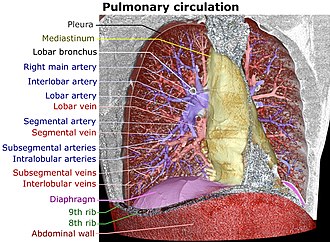
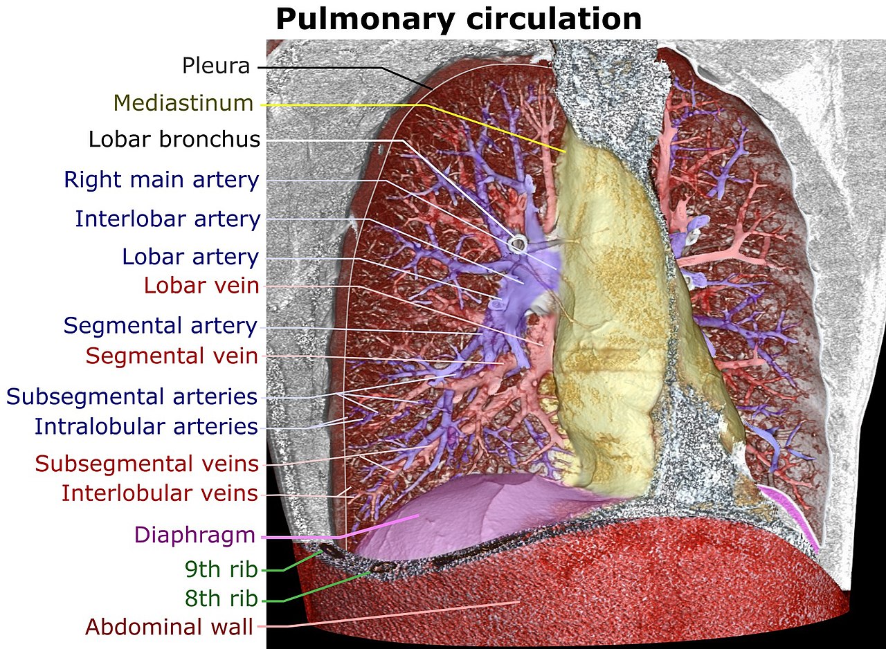
File:3D CT of thorax, annotated.jpg

本预览的尺寸:800 × 586像素。 其他分辨率:320 × 234像素 | 640 × 469像素 | 1,024 × 750像素 | 1,280 × 938像素 | 1,544 × 1,131像素。
原始文件 (1,544 × 1,131像素,文件大小:656 KB,MIME类型:image/jpeg)
摘要
| 描述 |
English:
|
| 日期 | |
| 来源 |
自己的作品
|
| 作者 | Mikael Häggström |
| 其他版本 |
|


许可协议
我,本作品著作权人,特此采用以下许可协议发表本作品:
| 本作品采用知识共享CC0 1.0 通用公有领域贡献许可协议授权。 | |
| 采用本宣告发表本作品的人,已在法律允许的范围内,通过在全世界放弃其对本作品拥有的著作权法规定的所有权利(包括所有相关权利),将本作品贡献至公有领域。您可以复制、修改、传播和表演本作品,将其用于商业目的,无需要求授权。
|
文件历史
点击某个日期/时间查看对应时刻的文件。
| 日期/时间 | 缩略图 | 大小 | 用户 | 备注 | |
|---|---|---|---|---|---|
| 当前 | 2017年6月25日 (日) 06:28 | | 1,544 × 1,131(656 KB) | Mikael Häggström | + Lobular |
| 2017年6月24日 (六) 19:34 |  | 1,644 × 1,062(632 KB) | Mikael Häggström | Corrected - there is very seldom a main pulmonary vein. Also, there's an interlobar artery segment on the right lung | |
| 2017年6月24日 (六) 14:37 |  | 1,644 × 1,062(639 KB) | Mikael Häggström | User created page with UploadWizard |
文件用途
全域文件用途
以下其他wiki使用此文件:
- ar.wikipedia.org上的用途
- bn.wikipedia.org上的用途
- en.wikipedia.org上的用途
- it.wikipedia.org上的用途
- ko.wikipedia.org上的用途
- ml.wikipedia.org上的用途
- ro.wikipedia.org上的用途
- sat.wikipedia.org上的用途
- simple.wikipedia.org上的用途
- vi.wikipedia.org上的用途